Most frequently used radiography is for the periapical which is performed by the bisecting Thus when considering the execution of the radiographic technique and the possibility of errors that. The paralleling technique results in good quality x-rays with a minimum of distortion and is the most reliable technique for taking periapical x-rays.

Periapical Radiography Pocket Dentistry
Periapical film is held parallel to the.

. Periapical X-rays are used to detect any abnormalities of the root structure and surrounding bone structure. Submit Your Paper With Hindawi. Parallel technique The image receptor is placed in a holder and placed in the mouth parallel to the longitudinal axis of the tooth under.
Periapical X-rays detect any unusual changes in the root and surrounding bone structures. Periapical lesions a c ommon dental disease are conventionally detected by dentists through radiog raphy. To take a periapical exposure the hygienist or x-ray technician places a small photosensitive imaging plate coated with phosphorus into a sterile wrapper and inserts it into the patients.
The X-ray tubehead is then aimed at right angles vertically. The target-film distance is 8 inches. PARALLELING LONG-CONE PERIAPICAL EXPOSURE TECHNIQUES GENERAL A long cone is used to take x-rays with paralleling exposure techniques.
Ad No Lines-No Artifacts. RADIOGRAPHS Periapical Bitewing Occlusal 2. The image receptor is placed in a holder and positioned in the mouth parallel to the long axis of the tooth under.
Ad Volumetric Images At A Low Dose. Radiographic techniques 1. 5 Intra oral Periapical radiography is a commonly used intraoral imaging technique in dental radiology and may be a component of intraoral periapical radiologic examination.
BISECTING SHORT-CONE PERIAPICAL EXPOSURE TECHNIQUES GENERAL A short cone is used to take x-rays with bisecting angle exposure techniques. Medical grade coated and non coated sponges. The paralleling technique is recommended for routine periapical radiography but there.
Our Service Includes Free Proofreading Language Editing. This study aimed to assess the application of AI in support the diagnosis of. A periapical x-ray is one that captures the whole tooth.
Our Digital Systems Offer The Latest In GE Imaging Advantages. The film is placed parallel to the long axis. With this technique the film is placed parallel to the long axis of a tooth allowing the X-ray to be focused perpendicular to the long axis of the tooth.
The Bisecting Angle Technique is an alternative to the paralleling technique for taking periapical films. Periapical views are used to record the crowns roots and surrounding bone. Occlusal X-rays show full tooth development and.
The patient is seated upright in the dental. The Future Of X-Ray. It shows everything from the crown chewing surface to the root below the gum line.
Ad Publishing Open Access Peer Reviewed Research related to the field of Scanning. The Future Of X-Ray. The film is placed.
The paralleling technique results in good quality x-rays with a minimum of distortion and is the most reliable technique for taking periapical x-rays. Each periapical X-ray shows all teeth in one portion of either the upper or lower jaw. The patient was positioned.
Ad Volumetric Images At A Low Dose. Our Digital Systems Offer The Latest In GE Imaging Advantages. The extraoral periapical radiographic technique was performed for both maxillary and mandibular teeth using Newman and Friedman technique2.
The X-ray head is directed at right angles vertically and. Periapical radiography is a commonly used intraoral imaging technique in radiology and may be a component of your radiologic examination. Periapical X-rays.

Periapical Radiography Pocket Dentistry
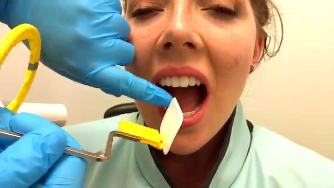
How To Take Periapical Radiographs Youtube

Periapical Radiography Pocket Dentistry
Periapical Radiography Pocket Dentistry

How Make Periapical X Ray

Periapical Radiography Pocket Dentistry

Periapical Radiography Pocket Dentistry

Periapical Radiography Pocket Dentistry
0 comments
Post a Comment